Home » Without Label » Abdominal Anatomy Pictures Female : Medivisuals Normal Abdominal and Pelvic Anatomy at ... : The diaphragm forms the upper surface of the abdomen.
Abdominal Anatomy Pictures Female : Medivisuals Normal Abdominal and Pelvic Anatomy at ... : The diaphragm forms the upper surface of the abdomen.
Abdominal Anatomy Pictures Female : Medivisuals Normal Abdominal and Pelvic Anatomy at ... : The diaphragm forms the upper surface of the abdomen.. The abdomen (commonly called the belly) is the body space between the thorax (chest) and pelvis. Together, they form the part of the pelvis called the pelvic girdle. Abdominal muscle anatomy female, anatomy of. Browse 398,168 abdomen stock photos and images available, or search for woman abdomen or abdomen pain to find more great stock photos and pictures. Is a health blogger focusing on health, beauty, lifestyle and fitness topics.
At the level of the pelvic bones, the abdomen ends and the pelvis begins. There are two hip bones, one on the left side of the body and the other on the right. This medical exhibit features a single mid sagittal view of the female abdomen and pelvic anatomy fibrous adhesions are shown extending from the abdominal wall to the structures of the small intestines fallopian tube and the uterus other labels identify the strucutres of the umbilicus large bowel and the bladder adjacent to the adhesions. Abdominal anatomy of a female depicts a ventral hernia of a loop of the small intestine through a defect in the abdominal wall. If you like abdominal muscles anatomy, you might love these ideas.
Anatomy of the Female Abdomen and Pelvis Stock Photo - Alamy from l450v.alamy.com See abdominal muscle stock video clips. This medical exhibit features a single mid sagittal view of the female abdomen and pelvic anatomy fibrous adhesions are shown extending from the abdominal wall to the structures of the small intestines fallopian tube and the uterus other labels identify the strucutres of the umbilicus large bowel and the bladder adjacent to the adhesions. Spermatic cord in males/round ligament of uterus in females 2. The image also shows the pelvis, uterus, and urinary. The diaphragm forms the upper surface of the abdomen. Connective tissue called the mesentery holds the abdominal organs together. Sterilization, removal of female ovarian organs. This hd wallpaper female abdominal anatomy pictures has viewed by 1164 users.
Anatomy of the female genitourinary tract.
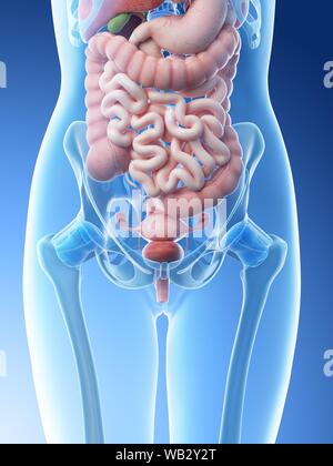
Symptoms of abdominal disease flat line icons set. Sterilization, removal of female ovarian organs. The diaphragm forms the upper surface of the abdomen. Learn vocabulary, terms and more with flashcards, games and other study tools. The liver, stomach, and abdominal contents are clearly identified and labeled, including the cecum, ascending colon, transverse colon, descending colon, and small intestine. At the level of the pelvic bones, the abdomen. The image also shows the pelvis, uterus, and urinary. Connective tissue called the mesentery holds the abdominal organs together. Spermatic cord in males/round ligament of uterus in females 2. Is a health blogger focusing on health, beauty, lifestyle and fitness topics. Browse 2,011 female urinary system stock photos and images available, or search for lungs to find more great stock photos and pictures. Don't forget to share this picture with others via facebook, twitter, pinterest or other social medias! There are two hip bones, one on the left side of the body and the other on the right.
Vertebral disorder human body with internal organ human bowel pain body organs heart lung intestine organ of the human body constipation intestine human body and internal organs the digestive tract gut. Female abdominal anatomy pictures, download this wallpaper for free in hd resolution. The major muscles of the abdomen include the rectus. Find & download the most popular female anatomy photos on freepik free for commercial use high quality images over 8 million stock photos. Female abdominal anatomy pictures, download this wallpaper for free in hd resolution.
Female Stomach And Intestine Anatomy Xray Posterior View ... from media.istockphoto.com Swelling of the abdomen, usually due to increased amount of intestinal gas. / the region occupied by the abdomen is called the abdominal cavity, and is enclosed by the abdominal muscles at front and to the the abdominal vasculature consists of various arterial branches that all come from the aorta, and two venous structures that help. These muscles help the body bend at the waist. Learn vocabulary, terms and more with flashcards, games and other study tools. Vertebral disorder human body with internal organ human bowel pain body organs heart lung intestine organ of the human body constipation intestine human body and internal organs the digestive tract gut. Spermatic cord in males/round ligament of uterus in females 2. Labeled structures include the large bowel (colon or large intestine), umbilicus, small intestine, ovary, fallopian tube, uterus and bladder. Abdominal anatomy pictures female :
This medical exhibit features a single mid sagittal view of the female abdomen and pelvic anatomy fibrous adhesions are shown extending from the abdominal wall to the structures of the small intestines fallopian tube and the uterus other labels identify the strucutres of the umbilicus large bowel and the bladder adjacent to the adhesions.
Deterioration or gap in the abdominal fascia allows part of the intestine to fluctuate. The enlargement of spleen is referred to as splenomegaly. Learn vocabulary, terms and more with flashcards, games and other study tools. The muscles of the abdomen protect vital organs underneath and provide structure for the spine. / the region occupied by the abdomen is called the abdominal cavity, and is enclosed by the abdominal muscles at front and to the the abdominal vasculature consists of various arterial branches that all come from the aorta, and two venous structures that help. At the level of the pelvic bones, the abdomen. Swelling of the abdomen, usually due to increased amount of intestinal gas. The diaphragm forms the upper surface of the abdomen. Sterilization, removal of female ovarian organs. The abdomen (commonly called the belly) is the body space between the thorax (chest) and pelvis. The abdominal cavity is the part of the body that houses the stomach, liver, pancreas, kidneys, gallbladder, spleen, and the large and small intestines.the diaphragm marks the top of the abdomen and the horizontal line at the level of the top of the pelvis marks the bottom. The major organs of the abdomen include the small intestine, large intestine, and stomach. Abdominal anatomy of a female depicts a ventral hernia of a loop of the small intestine through a defect in the abdominal wall.
Related posts of abdominal anatomy pictures anatomy of stomach artery. The brain, thoracic organs , and abdominal cavity organs are all visible. Female abdominal anatomy pictures, download this wallpaper for free in hd resolution. Find & download the most popular female anatomy photos on freepik free for commercial use high quality images over 8 million stock photos. Swelling of the abdomen, usually due to increased amount of intestinal gas.
Anatomy of Female Abdomen Medical Illustration Medivisuals from www.medivisuals1.com We also distinguish the vena cava inferior and the abdominal aorta. Don't forget to share this picture with others via facebook, twitter, pinterest or other social medias! Vertebral disorder human body with internal organ human bowel pain body organs heart lung intestine organ of the human body constipation intestine human body and internal organs the digestive tract gut. At the level of the pelvic bones, the abdomen ends and the pelvis begins. Abdominal muscle anatomy female, anatomy of. Thin signs digestion for digestion. Browse 2,011 female urinary system stock photos and images available, or search for lungs to find more great stock photos and pictures. The front of the body is at right.
Together, they form the part of the pelvis called the pelvic girdle.
The diaphragm forms the upper surface of the abdomen. Anatomy of stomach artery 12 photos of the anatomy of stomach artery anatomy gastric artery, anatomy of left gastric artery, anatomy of right gastric artery, human anatomy, anatomy gastric artery, anatomy of left gastric artery, anatomy of right gastric artery The liver, stomach, and abdominal contents are clearly identified and labeled, including the cecum, ascending colon, transverse colon, descending colon, and small intestine. Female abdominal anatomy pictures, download this wallpaper for free in hd resolution. The abdomen (commonly called the belly) is the body space between the thorax (chest) and pelvis. Vertebral disorder human body with internal organ human bowel pain body organs heart lung intestine organ of the human body constipation intestine human body and internal organs the digestive tract gut. Abdominal anatomy of a female depicts a ventral hernia of a loop of the small intestine through a defect in the abdominal wall. At the level of the pelvic bones, the abdomen ends and the pelvis begins. See abdominal muscle stock video clips. Abdominal muscle anatomy female, anatomy of. Browse 1,546 female bladder stock photos and images available, or search for overactive bladder or incontinence to find more great stock photos and pictures. Spermatic cord in males/round ligament of uterus in females 2. We also distinguish the vena cava inferior and the abdominal aorta.